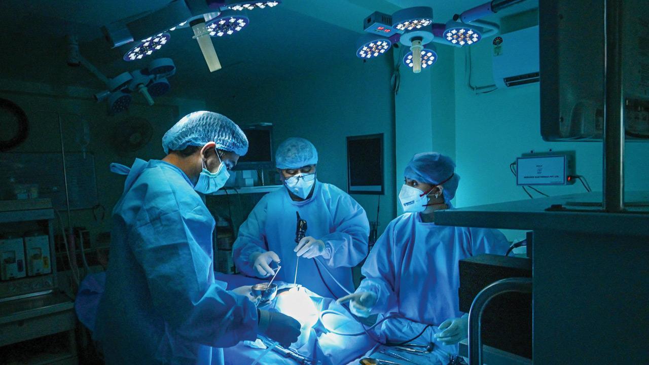
Overconfidence at the operation table need not always translate into a successful surgery; it’s the unpredictability that keeps most doctors on their toes

Representative Image
 Mrs Patil walked into the clinic with her daughter. She was in her 60s, wore a hand-woven saree, and tied her hair in a bun. She was the principal of a renowned school and rested her glasses low on her nose to complete the look. When she spoke to me, her eyes peered above her spectacles, taking me back to my school days, when our teachers glared at us when they wanted to appear to be strict. Unfortunately, she had a problem with her vision. “I am unable to see on my sides,” she mentioned. “When I’m attempting to cross a road, I can’t tell if a car is coming, and I keep bumping into things in the house,” she went on to explain.
ADVERTISEMENT
Her eye doctor was quick to realise that this was a sign of optic nerve compression and asked for an MRI of the brain, which her daughter pulled out amidst sheaves of other reports. After I examined her, I wedged the films into the viewing box and went on to explain what we all knew. “She has a gigantic tumour arising from the pituitary gland in the centre of the head that’s grown large enough to push the optic nerves up. Both sides of her visual fields are affected because the tumour is compressing the junction of both optic nerves as they leave the eye,” I tried to explain simply.
“Will you be able to remove this from the nose,” she asked point blank, obviously having done her research and taken a few opinions before coming to me—because the other option is to excise it by sawing the head open. This time I peered at her with eyes above my glasses. “I don’t want to open up my head,” she said categorically. “It’s possible to remove this from the nose,” I said after studying the MRI more carefully, “but it’ll be slightly risky,” I confessed, showing her a part that was probably stuck to an artery and another part that had extended beyond the line of vision. “We’ll have to do an expanded endonasal approach,” I said, to make it sound fancy. I explained the risks of death, paralysis, coma, infection, bleeding, and hormonal imbalance, and saw her complexion turn ashen. I quickly reassured her that more often than not, everything goes well. Until it doesn’t, I said to myself.
The human nose is one of the most fascinatingly designed parts of our anatomy. The Parsi nose of course, does not fit this description. But, irrespective of gender or race, when you look at the nose through an endoscope, it is visually spectacular—once you’ve gotten past the hair in it. Entering the nose to reach the brain is like trekking from Manali to Leh: the turbinates, with their shades of pink and brown, resemble the rugged valleys and mountainous terrain of that scenic route. Once we went pass those, we enter the gaping sphenoid sinus, its topography changing completely to something more cavernous, with thin bony septa guiding us up further to our destination. Just as trekkers find it adventurous to identify various peaks around them while climbing, surgeons like to do the same with anatomy. We like to demonstrate to everyone in the operating room where we are in our journey to our final destination, and in doing so, we reached the widened floor of the sella, a saucer-shaped bony structure that housed the pituitary tumour and was thinned out by it. I took a diamond drill and caressed it as bone turned into dust, which we washed away. I then drilled a little more ahead so we had access to the extensions of the tumour. We made enough room for us such that we could operate through both nostrils using four hands, two of mine and two of my ENT colleague’s. I cut into the bottom of the tumour and it was soft and gooey, like they most often are in this location. Using a suction in one hand and a curette in another, I kept gently dislodging the tumour and allowed it to descend into the cavity we had created.
The challenge was to get to the part that had insinuated itself into the frontal lobe of the brain. It was harder, not as mushy as the rest of it, and I had to hold it with forceps and gently tease around it to free it up from spiderweb-like strands to remove it completely. Just as the last bit got extricated, there was a sudden onset of torrential bleeding and the nose filled up with blood like a volcanic eruption, blurring the lens of the endoscope. It was akin to a trekker slipping off the top of the summit and rolling down the mountain, splattered in blood. I put in a much larger suction, got the anaesthesiologist to lower the blood pressure, and briskly identified a small twig that was bleeding, buzzing it with a bipolar cautery.
Three thousand thoughts must have crossed my mind in the three minutes it took to control this. The scenic vistas, temporarily marred by what looked like a forest fire, were once again restored to their pristine glory. The brain was soft and pulsating gently, the optic nerves free from what was burdening them. We closed meticulously in the usual fashion, layering the base of the skull to seal the defect from below. I walked out of the operating room with broad shoulders, tapping myself on them for having performed a great operation.
She woke up with completely restored vision, which was a miracle in itself, and was transferred out of the ICU the next morning. A few days later, she was sitting up in her bed, hair oiled back, spectacles low on her nose, reading the newspaper. She looked at me from above her glasses as I walked in to meet her. Neither of us could believe she had a massive brain tumour surgery just a few days ago. I explained to her how modern surgery had evolved, and that with improvements in technology and visualisation, we are now able to do what was previously the unthinkable. She was discharged after a week once everything was in check. Not only did I walk about with broad shoulders, I pumped my chest out a bit as well. This is how most neurosurgeons routinely walk.
But medicine, as we know time and again, is a great leveler. Two weeks later, I got a call from the ER saying she had returned with severe headache and vomiting. While she was conscious when she came in, she was no longer responding within the next few minutes. A CT scan, which had been clean when she was discharged, now looked like a bomb had burst in her head. An angiogram was not able to reveal the source of the bleed. I suspect that the twig we had coagulated had probably opened up again, but I wasn’t able to prove it. Over the next few weeks, we got her off the ventilator as she showed some signs of improvement and was on her way to recovery that was slow, but sustained.
The case that had made me want to show off my surgical skills a while ago had made me want to give up operating a few weeks later. The case that had showed me breathtaking views of the mountains only a few days ago, had them crumble and collapse on me in an instant. The case that had showed me the light to be able to push the boundaries of modern medicine, had, within a matter of time, swallowed it up into a silent scream of darkness. The nose, which until some time ago was a stairway to heaven, had turned out to be a highway to hell. Such is life…
The writer is practicing neurosurgeon at Wockhardt Hospitals and Honorary Assistant Professor of Neurosurgery at Grant Medical College and Sir JJ Group of Hospitals.
 Subscribe today by clicking the link and stay updated with the latest news!" Click here!


